




| IN A NUTSHELL |
|
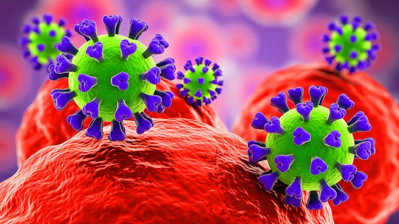
The return of winter often brings with it the common symptoms of flu: fever, aching limbs, and a runny nose. Influenza viruses are the culprits behind these ailments, entering the body through droplets and infecting susceptible cells. A groundbreaking study by a team of researchers from Switzerland and Japan has provided an unprecedented glimpse into the behavior of these viruses. Using a novel microscopy technique, they have been able to observe, in real-time, the moment an influenza virus infiltrates a living cell. This discovery sheds new light on the intricate dance between viruses and human cells, offering potential pathways for future antiviral research.
Viral Surfing on the Cell Surface
The influenza virus begins its infection process by attaching to specific molecules on the surface of a human cell. This action is quite similar to surfing, where the virus glides along the cell membrane, latching onto one molecule after another. This journey leads it to a spot densely populated with receptors, which serves as the most efficient entry route. The infection process appears active, as the virus hijacks a routine cellular uptake system. This system, essential for the cell’s survival, typically brings in hormones, cholesterol, and iron.
Once the virus attaches to the cell’s receptors, the membrane starts forming a small indentation at the point of contact. A structural protein called clathrin supports and shapes this deepening pocket. As the pocket grows, it envelops the virus, forming a vesicle. The cell then pulls this vesicle inward, where the coat dissolves, releasing the virus into the cell. This process, while seemingly simple, represents a complex interaction that the virus has mastered to ensure its survival and proliferation.
Jaya Anand Singh’s Research Path : A Journey from Curiosity to Contribution
Why Earlier Microscopy Fell Short
Previous attempts to capture the critical moment of viral entry into cells faced significant limitations. Techniques like electron microscopy, although detailed, required the destruction of cells to capture images, resulting in mere snapshots of the interaction. In contrast, fluorescence microscopy allowed for live imaging but suffered from low spatial resolution. These shortcomings left gaps in understanding the precise mechanics of viral entry.
The advent of the new microscopy approach, ViViD-AFM, marks a significant leap forward. By combining atomic force microscopy (AFM) with fluorescence microscopy, researchers can now track the virus’s fine-scale movements as it enters the cell. This dual approach provides unprecedented clarity and detail, allowing scientists to observe the dynamic process as it unfolds in real-time.
ViViD-AFM Sheds Light on Viral Entry
The introduction of ViViD-AFM has revolutionized the study of viral entry into cells. This advanced tool revealed that cells play an active role in assisting the virus at several stages of entry. The cell’s membrane pushes upward, seemingly trying to capture the virus. Additionally, clathrin proteins are summoned to the site of attachment, further facilitating the virus’s entry. These wave-like motions intensify if the virus attempts to drift away from the surface, suggesting a highly coordinated interaction.
ViViD-AFM’s ability to provide real-time visualization has significant implications for antiviral research. By offering a detailed view of the infection process, it allows scientists to test antiviral drug candidates directly in cell cultures. This method could also extend to studying other viruses or vaccines, providing a new perspective on how these particles interact with human cells.
Implications for Antiviral Research
The real-time insights offered by ViViD-AFM pave the way for breakthroughs in antiviral research. This technique enables scientists to observe the infection process as it happens, offering a valuable platform for testing potential antiviral drugs. The implications extend beyond influenza, as the method can be adapted to study other viruses or even vaccines. Understanding the precise mechanics of viral entry can lead to the development of more effective treatments and preventive measures.
Moreover, this research underscores the importance of cellular interactions in the infection process. By highlighting the active role cells play in facilitating viral entry, new targets for antiviral interventions may be identified. The detailed observations made possible by ViViD-AFM could revolutionize how researchers approach the study of viral infections and the development of corresponding therapies.
As the world continues to face challenges posed by viral infections, the insights gained from this research highlight the critical role of advanced microscopy techniques in understanding these complex interactions. With ViViD-AFM, scientists are equipped with a powerful tool to delve deeper into the mechanics of viral entry. Could this breakthrough lead to a new era of antiviral treatments that effectively halt infections before they take hold?



Wow, this sounds like a game-changer for flu research! 🧬
Wow, this ViViD-AFM technique sounds like something out of a sci-fi movie! Incredible work by the scientists! 🚀
Can this technique be used to study COVID-19 as well? 🤔
Is this technique applicable to other viruses like COVID-19? 🤔
Incredible work by the researchers! Thank you for sharing this breakthrough. 🙌
Finally, some good news in science! This could be a game-changer for flu season. Thank you to the researchers involved! 🙌
The surfing analogy was a nice touch. Makes the process more relatable!
Why did it take so long for a technique like ViViD-AFM to be developed? It seems like a huge leap forward.
This is great, but how long before we see new treatments as a result?
How accessible is this new microscopy technique for labs around the world? Will it be widely used?
ViViD-AFM sounds like science fiction brought to life. Amazing!